ORAL SURGERY · WARSAW
Dental and jaw cysts
in Warsaw
A change seen on an X-ray needs diagnosis and assessment of its relationship to teeth and nearby structures before treatment is chosen.
Not every radiolucent change is a cyst and different cysts are managed differently. The plan follows examination, imaging and, when indicated, tissue assessment.
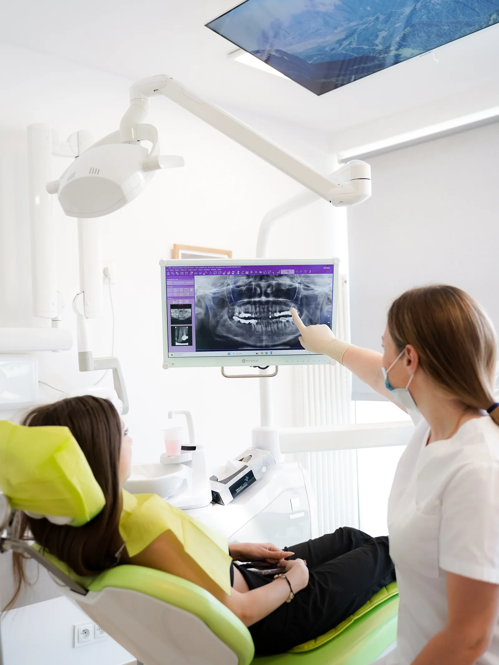
01Diagnosis firstA procedure name should not replace identification of the lesion.
02Location shapes treatmentSize and proximity to teeth, nerves and the sinus can change the approach.
03Tissue may need examinationHistopathology can help establish the diagnosis and follow-up.
01 / CONSULTATION
When is a surgical consultation useful?
The starting point may be a symptom or an unexpected finding on imaging taken for another reason.
- 01
A change reported on an X-ray
We clarify what the description means and whether more diagnostics are needed.
- 02
Repeated swelling or discomfort
Symptoms require diagnosis rather than self-identification of a cyst.
- 03
A change near a previously treated tooth
The tooth, bone and possible source are assessed together.
- 04
A referral to oral surgery
We organise the reason for referral, available records and possible next steps.
02 / STAGES
From identifying the lesion to follow-up
The sequence varies according to the suspected origin, symptoms, size and position.
- 01
History and examination
We ask about symptoms, previous treatment and health.
- 02
Imaging and differential diagnosis
Available images are reviewed and further assessment is selected only when needed.
- 03
Treatment options
Observation, treatment of a related tooth, decompression or surgical removal may be discussed depending on the diagnosis.
- 04
Procedure and tissue examination
When a lesion is removed, the material may be sent for histopathological assessment.
- 05
Results and follow-up
The diagnosis and healing determine the review schedule and any further care.
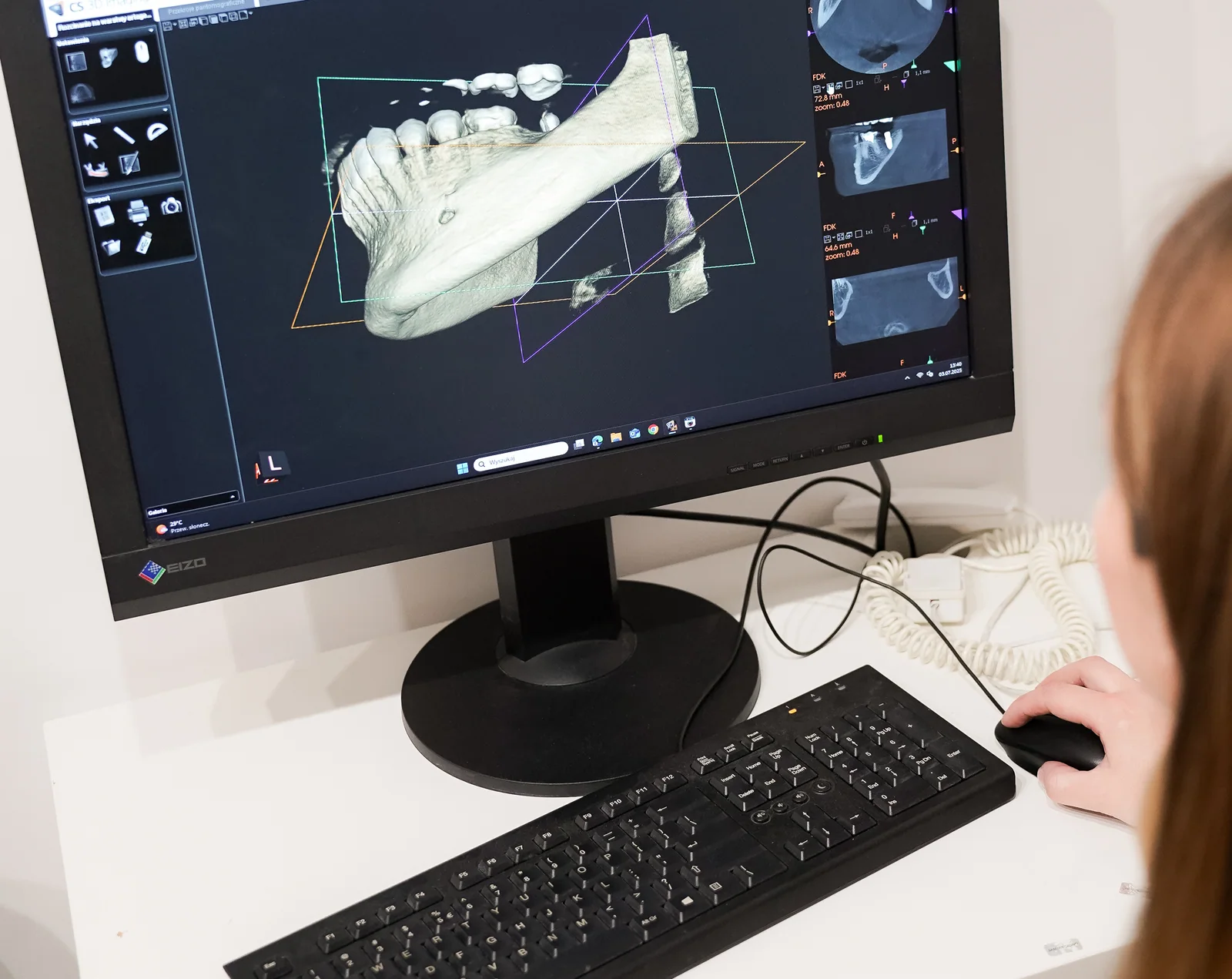

03 / INDIVIDUAL PLAN
What determines the treatment plan?
Size alone does not determine management. Origin and nearby anatomy are equally important.
Likely type and origin
Dental and non-dental lesions can require different management.
Size and extent
A larger lesion may need a staged or decompressive approach.
Nearby structures
Teeth, nerves and the sinus are considered in planning.
Health and healing
Medical history and medicines affect preparation and aftercare.
04 / BEFORE YOU DECIDE
What do we discuss before treatment?
- the report and images that first identified the change;
- previous treatment of teeth in the area;
- current symptoms and how they have changed;
- possible approaches and the reason for tissue examination;
- how and when results and follow-up will be provided.
Important: this page provides general information and does not replace an examination or individual consultation. Suitability and the final plan are established after clinical assessment.
05 / PATIENT QUESTIONS
Common questions before a consultation
Questions about your symptoms or imaging are best discussed during an individual appointment.
Is every change on an X-ray a cyst?
No. Different conditions can look similar, which is why diagnosis comes before treatment.
Does every cyst need immediate surgery?
No. Management depends on diagnosis, size, symptoms, location and possible alternatives.
Why might tissue be sent to a laboratory?
Microscopic examination can confirm the nature of the removed tissue and guide follow-up.
Can a related tooth be preserved?
Sometimes. The prognosis of the tooth and its relationship to the lesion must be assessed individually.
Will the cyst come back?
Recurrence risk depends on the diagnosis and treatment, so the follow-up plan is specific to the pathology result.
MEDICAL SOURCES
Patient information used for review
this page provides general information and does not replace an examination or individual consultation. Suitability and the final plan are established after clinical assessment.
FIRST STEP
Let’s start with your situation.
Choose a practice, check available appointments and book a consultation.